 This webcast features:
Dr. Josefina Nilsson, Head of Vironova Services, as well as her colleagues, Gustaf Kylberg and Mathieu Colomb-Delsuc.
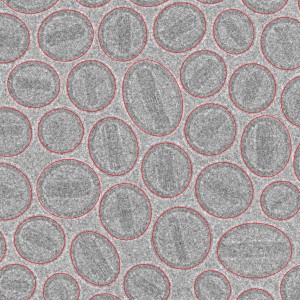
Advanced analytics that provide objective and reliable data can help to improve processes and shorten development time of drug and gene delivery platforms. The use of electron microscopy (EM) imaging combined with analysis using the proprietary Vironova Analyzing Software (VAS) enables semi-automated particle detection and classification. Statistically significant results are obtained in a time and cost effective manner. Typical data includes size distribution, level of packaging, individual particle morphology, thickness of the lipid bilayer, lamellarity and presence of debris. This type of information reveals of prime importance when performing drug characterization analysis for encapsulated drugs, as these characteristics not only reflect on the quality of the development and manufacturing processes, but also influences biological activity, bio-distribution, and toxicity.
During this webinar, Dr. Josefina Nilsson presents how EM imaging together with VAS analysis can provide objective characterization data of liposomal drug carriers and viral vectors in a time and cost efficient way.

I am interested in the capability to determine whether or not a set of particles are filled or empty. May I know in more details how this works, and if I have a sample, what can I do to obtain this capability?
Regards,
Anastasia