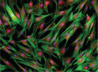
Biopreservation suppresses degradation and enables postpreservation recovery of structure, viability, and function. Although there are several biopreservation techniques (indicated in “Biopreservation Methods” box), most laboratories use either standard cryopreservation protocols (the far majority) or vitrification (much more limited in broad systems application) when freezing cells for research and clinical applications. Isopropanol freezing containers such as the Mr. Frosty device from Nalgene Labware have made cryopreservation easier in many applications, and controlled-rate freezers allow users to program and manipulate freezing rates.
Nonetheless, cryopreservation is not often given the same attention as other bioprocessing steps. I discussed these issues with Aby J. Mathew, a noted biopreservation expert and senior vice president and chief technology officer at BioLife Solutions. His opinion is that developers of cell therapy and regenerative medicine products often focus first on cell characterization, method of action, and potency assays, and allow cryopreservation (or any biopreservation processing steps) to become the weak link in their bioprocessing work f low chain. “If you put a lot of time and effort into your cell therapy and then just freeze it down into a freeze media that you either make in-house or buy from a supplier, are you actually spending the same amount of time and energy making sure that your cryopreservation process is actually just as strong as the rest of your system?”
Mathew says that cell therapy groups often do not send out in-house freeze media for testing nor follow robust release criteria for the freeze media. “They might follow an SOP, but it is not necessarily followed as a validated batch record.”
An Optimized Approach
Sensitivity to biopreservation stress depends on cell type. Methods can be optimized by changing freeze and storage media formulations and freeze–thaw protocols. Many cell therapy groups use the most streamlined and consistent processes and do not necessarily want to invest time in optimizing freeze media or cooling rates, says Mathew. “Keep in mind that process development folks tend to be engineers or manufacturing personnel, not ‘basic scientists’ who want to make cryopreservation a research project. Many of my colleagues in cryobiology develop mathematical models, and study the physics of the cell freezing process. Although not the only group, our team’s approach was different in that we built off these fundamental principles that had been developed for several decades, and then added some newer molecular biology elements to this previous body of knowledge.”
Rather than looking at cells from the outside in, Mathew and colleagues approached it from the inside out (1,2,3). “We isolated DNA, RNA, proteins, and so forth from cells following cryopreservation and hypothermic preservation (2–8 °C), and observed that apoptotic signaling/death pathways were initiated in addition to necrotic pathways. We also saw elevated stress markers, such as free radical generation. Our approach was not only to determine how to ‘buffer’ cells from the stresses that contribute to cell death but also to reduce stress on surviving cells.”
Engineered cryopreservation media such as the CryoStor brand and home-brew cocktails such as culture media with the cryoprotectant dimethyl sulfoxide (DMSO) can work adequately for a number of cell types. However, postthaw viabilities may vary because cells have different sensitivities to the preservation-induced stress. Some freeze media can be used uniformly. “We know that some customers have qualified and validated our CryoStor their internal banking system for a number of cell types. Others will take a culture media for specific cells then add DMSO — with or without serum or protein — and define that as their cryopreservation media for those specific cells. In other words, if the culture media was optimized for specific cells, then the view by some is that media should be part of the freeze media.” Product sheets for some cell suppliers, for example, will indicate cryopreservation of cells in a specific culture media plus 10% DMSO. Such approach may create additional labor (and potential increased risks), says Mathew. “This would require formulation of a minibatch of freeze media specific to X cell type, and a separate, differently formulated minibatch of freeze media specific to Y cell type.”
Equipment
One impediment to broader awareness of best practices says Mathew, is that many commercial companies are developing improved manufacturing systems, but do not publish or share their knowledge. That may not be a hindrance for most cell therapy groups that are developing cellular products on a smaller scale. Those groups are able to get by with using isopropanol containers or small-scale controlled-rate freezers for freezing cells in vials before storing them into liquid nitrogen dewars.
BIOPRESERVATION METHODS
Not all of the methods of biopreservation are broadly validated into cell therapy manufacturing. Biopreservation could include ambient storage, hypothermic storage (2–8 °C generally), standard cryopreservation, vitrification (ice-free cryopreservation), and anhydrobiosis (drying or desiccation). For most clinical applications, you usually see the application of hypothermic preservation (if a nonfrozen application is used) or standard cryopreservation (generally a DMSO concentration <20% in some vehicle media solution with a freezing rate of ~–1 °C/min).
One measure of the broad use of these applications is evaluating solutions that can be found in laboratory catalogs. “For the most part you will find hypothermic preservation solutions (or transport media), and standard cryopreservation freeze media along with freezing devices and similar generic liquid nitrogen dewars and controlled-rate freezers. The broad acceptance of a method or its incorporation into validated protocols by ‘nonexperts’ is probably an indicator of its utility, especially within process development applications.”
However, cell therapy groups and commercial companies moving into larger scale production to support later stage clinical trials and commercial shipments will likely need further bioprocessing optimization. “Instead of working with cells in flasks that are centrifuged into a clean pellet from the culture media, they might be working with cells in cell factories or bioreactors where cells might be concentrated down to remove the majority of liquid so that the cells are left in a slurry. Cryopreservation media is added to this slurry and then cells are siphoned off into different aliquots and packaged into large vials or large bags for freezing. The optimization of this entire process is rarely shared publicly due to commercial confidentiality interests and/or the lack of ‘sexiness’ for acceptance as an oral presentation at a scientific conference.”
Testing
One current debate focuses on determining the appropriate assa
ys to ensure cell potency or functionality. There are no standard testing methods to ensure cell viability, and most procedures resemble those used for cell culture, even though they are not always applicable. For cell therapy, viability is often assessed immediately postthaw by simple live–dead assays that may not indicate the true, long-term viability of the cells because they do not address the phenomena of delayed-onset cell death. However, in a clinical application in which preserved cells are injected or infused into patients, there may not be the luxury of monitoring the cells extensively at that point in the process. This is where the need for appropriate validations of a cell or tissue product and its manufacturing controls demonstrate the strength of a good system.
“What you are trying to determine is whether the same potency remains over the period of time between when a cell therapy is manufactured and when it is applied to a person,” says Mathew. “If you were having a bone marrow stem-cell transplant in a local hospital, your bone marrow stem cells would usually be frozen down and then thawed out. The laboratory would perform a simple trypan blue assay — a pass or no-go checking point. As long as that gives them high enough numbers, they move forward. But the question we bring up many times is that if you tested this right after it was thawed, how do you know two hours or even 12 hours later, that those cells are actually surviving — especially after they are infused into a patient?”
Current methods may not necessarily provide that information, and companies might want to make this information part of their premanufacturing validations, says Mathew. “In that way, you could say that because you’ve tested the system so many times in a more stringent manner, you know that those cells are still going to be okay.”
Even if this extended testing is not conducted, cell therapy manufacturers must still validate that the cell product is the same as what was previously characterized (within their release criteria). If the cell product meets potency assay requirements, and acceptable levels of viability and functionality are observed, then the postpreserved product could be considered equivalent. “In addition, developers have to qualify all of the reagents that the cells are exposed to,” says Mathew. “From a commercial perspective, this is part of the reason our company developed fully defined solutions that are serum-free and protein-free — they integrate well into the quality and regulatory systems of cell therapy manufacturers. Academic and R&D settings often use culture media with serum and DMSO to freeze cells. “The use of undefined serum components in freeze media for clinical applications may pose additional risk and could require additional risk analysis, validation, and costs.”
Media Validation
Best cryopreservation practice in GMP clinical applications includes the use of fully defined, serum-free and protein-free, intracellular-like cryopreservation media; similar to the advances made in organ transplant preservation media formulation 25 years ago. Another biopreservation or bioprocessing optimization consideration relates to minimal manipulation at the clinic. A decision must be made to centrifuge and wash off the cryopreservation media, dilute the final product with infusion-grade solution, or use the freeze media as the excipient vehicle and/or delivery solution for the cell therapy product (meaning it is not removed and is infused into the patient). “Cell therapy developers and clinicians should qualify the benefits of not losing or damaging cells through a wash step in this excipient application” says Mathew. In this situation, more scrutiny is needed on the qualifications of the cryopreservation media, or equivalent preservation media if it is a nonfrozen cell therapy product. Developers and clinicians have a responsibility to qualify and validate excipient reagents within their system and specific application.”
Mathew’s company helps their customers design appropriate experiments to qualify biopreservation media for an intended use. To facilitate the process, the company’s products are supported by FDA master files. Mathew offers this final thought, “The ultimate goal of the corporate and nonprofit cell therapy and regenerative medicine markets is the development of cell, gene, and tissue-based products and therapies that offer the maximum therapeutic benefit with the least risk in the most cost-effective manner for the patient, payers, and manufacturers. It is the responsibility of each of us involved in this process to develop as fully-optimized processes as possible.”
Author Details
Maribel Rios is managing editor of BioProcess International, mrios@bioprocessintl.com
REFERENCES

